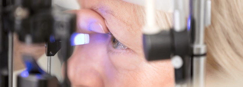
Comprehensive Eye Examination

iStock 666923606

A comprehensive eye exam will evaluate not only how well you see, but also identify potential eye diseases. Some eye diseases, such as glaucoma and macular degeneration, can result in serious vision loss if not detected and treated early. Often patients with these diseases don’t experience any visual symptoms before vision loss occurs.

If you are over 40, you should have a comprehensive eye exam every year, especially if you have family history of glaucoma, diabetes or diabetic retinopathy.

What To Expect At Your Eye Exam:

Your doctor will most likely dilate the pupils of your eyes, in order to better see the retina at the back of your eye. You may want to consider making transportation arrangements, as your vision may be blurry for a few hours after dilating.

Your Exam May Include The Following:

  • Visual acuity or refraction test to determine the degree to which you may be nearsighted, farsighted or have astigmatism
  • Muscle function test to check the movement of your eyes in each direction and at specified angles. This test will identify any muscle weakness or involuntary eye movement.
  • Binocular vision skills assessment to ensure that your eyes work together properly as a team. This is important for proper depth perception, eye muscle coordination and the ability to change focus from near to far objects.
  • Visual field test to measure your peripheral vision, the width of the area you can see when you’re looking straight ahead. This test may also detect diseases of the eyes or neurological disorders.
  • Eye pressure test. Your doctor may administer one or more tests to evaluate your intra-ocular pressure. High intra-ocular pressure may be a sign of glaucoma.
  • Color vision screening to see if you perceive colors properly.
  • Eye health assessment using an ophthalmoscope. This tool allows the doctor to evaluate your pupil responses, optic nerve, retina, cornea and lens.

Treatment options, if necessary will be presented at the conclusion of your examination.

Frequently Asked Questions:

Farsightedness (hyperopia) occurs when a patient’s eyes are too short for the curve of their cornea. As light rays enter the eye through the cornea they change direction or are refracted as they travel to the retina. If the light focuses behind the retina because of the cornea’s shape, the image in the brain can be blurred not only for objects at a distance but also up close. To know whether or not you are farsighted you’ll need a refraction examination performed by a trained optometrist or ophthalmologist.

With nearsightedness (myopia), the cornea’s shape causes light rays to focus in front of the retina. As a result nearsighted patients can see clearly up close but have blurry vision to the side and at a distance. To know whether or not you are nearsighted you’ll need a refraction examination performed by a trained optometrist or ophthalmologist.

Presbyopia is generally a “40 thing” – a consequence of aging. Normally, you have eye muscles that change the shape of the lens to focus on objects near to you. Around age 40 to 45, these muscles may become weak and not work as well with up-close objects as before. People experiencing this will begin pushing things away to see them more clearly. It’s also the time when people begin wearing magnifying or “reading” glasses” or some form of bi-focal-progressive glasses or contacts for correction.

There are a couple of ways to check for astigmatism, an abnormal shape of the cornea. We can perform a test called corneal topography to map out and examine the precise shape of the cornea and whether it contains any areas of steepness, flatness or other abnormalities. If we find a bow-like shape with one side of the cornea steeper than the other side, this can cause the light rays coming into the eye to focus on different parts of the retina depending on which direction they have entered.

Another method is to perform a refraction examination to check a patient’s vision prescription. We calculate measurement numbers for correcting the vision based on what the patient is seeing during the exam, rather than viewing the actual shape of the cornea. The results of a refraction examination can indicate the presence of astigmatism.

There are a number of ways to correct astigmatism. Traditionally, we can prescribe glasses or contacts as with other refractive problems. In recent years, refractive surgeries, particularly laser-based LASIK, may be used to correct astigmatism. And depending on the type of lens implant (IOL) used to replace a clouded natural lens we may be able to correct astigmatism as a result of cataract surgery.

Keratoconus is a particular form of astigmatism. Patients with this condition often have a steep angle to the shape of their cornea (often called a “beer belly” cornea). That angle changes the direction (refract) of the light traveling through to the retina in such a way that it’s sometimes difficult to correct with a vision prescription. Many patients don’t know they have this type of astigmatism unless they’re examined, corneal topography being one of the best ways to accurately diagnose this condition. Not only is it difficult to obtain an accurate prescription with keratoconus but LASIK surgery could make the condition worse.

Most people with normal vision will be able to see an object clearly at 20 feet. Using this as a benchmark, we ask a patient during an eye exam to view a chart of letters, numbers or symbols positioned about twenty feet away. If they can see a certain line they are considered “20/20,” meaning they have normal vision. If they can’t, but can see larger lines above it, we may calibrate that as 20/25, 20/30, and so on.

If a person can’t see 20/20, we must then consider if they have a refractive error (correctable with glasses, contacts or refractive surgery like LASIK), or another problem like cataracts or impairments of the retina or optic nerve. Because there are a number of possible causes to less than normal vision, a person should undergo a comprehensive exam to determine the exact cause.

Because of a structural issue with the eye, such as a condition like macular degeneration or legal blindness, we may not be able to correct their vision with glasses or similar means. We term this low vision. Patients with low vision can’t see clearly enough to read, watch television or perform other activities that require clear vision. We may refer such patients to a low vision aid specialist to explore other ways of improving their vision and quality of life.

Monovision is a way to help individuals that are both presbyopic, farsighted and myopic, nearsighted. With a monovision technique we can designate one eye for distant viewing (usually the dominant eye) and design a contact lens specifically for distance. We then designate the other eye for near vision and design a lens appropriate for that eye.

We can also achieve the same result with cataract surgery. We replace the eyes’ natural lenses with artificial implants known as intraocular lenses (IOLs). We fashion an IOL for distance in one eye and an IOL for near vision in the other eye.

Monovision is usually preferred by patients who’ve previously worn contact lenses but don’t wish to use reading glasses to correct their presbyopia. This method does affect depth perception to a small degree but the brain can compensate for that in other ways. Most patients are happy to have another alternative to glasses.

Pink eye (conjunctivitis) is an infection of the eye caused by an adenovirus. It typically begins in one eye becoming matted and turning red in the morning, followed in a couple of days with the other eye becoming infected. During an eye exam we may also observe other inflammatory reactions including follicles in the conjunctiva or white dots on the cornea.

A person with pink eye-like symptoms should undergo an eye exam and not attempt to treat themselves. Although the infection usually runs its course an eye physician may place a patient on antibiotics or steroids to help with healing. We also want to rule out a bacterial infection as the cause.

Strabismus is a condition in which one eye turns in, out, up or down differently from the other eye. It can be caused by a number of things: for example, if it occurs early in life it may be that the child’s eyes have different vision prescriptions and the dominant eye is overriding the other eye in order to focus. In older patients strabismus may result from strokes in the blood vessels of the eye that cause certain eye muscles to no longer work properly. It can also be related to high blood pressure or diabetes in which the nerves related to specific muscles become compromised. The affected eye can no longer function properly and thus no longer turn in tandem with the other eye.

With any case of strabismus, it’s important to undergo an eye exam with an ophthalmologist to determine the exact cause. This will help guide the physician to determine the best treatment approach.

Get In Touch With Us:

To learn more about Hattiesburg Eye Clinic and our services please call us at 601 268 5910 or toll-free at 800 624 8254 or schedule a consultation.

Book An Appointment

* All indicated fields must be completed.
Please include non-medical questions and correspondence only.

Location Map:

Accessibility Toolbar